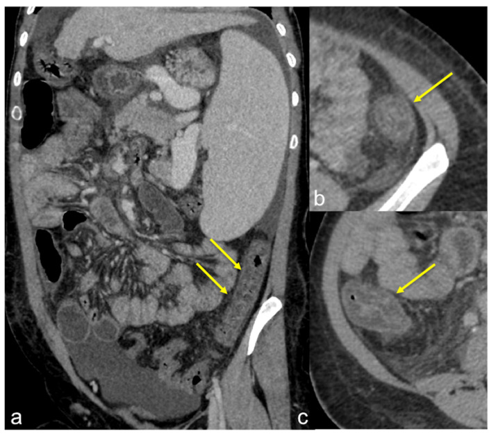
Figure 41.
Ischaemic colitis in a cirrhotic patient. Enhanced CTA scan in portal venous phase in coronal-oblique (a) and axial (b,c) views. Note the typical colonic involvement (a–ca;b;c;b;c;d;d;e;f;d;e;f;a;b;c;a;b;c;d) arrows) with stratified wall thickening with submucosal oedema, mucosal hyperaemia and adjacent mesenteric stranding.

