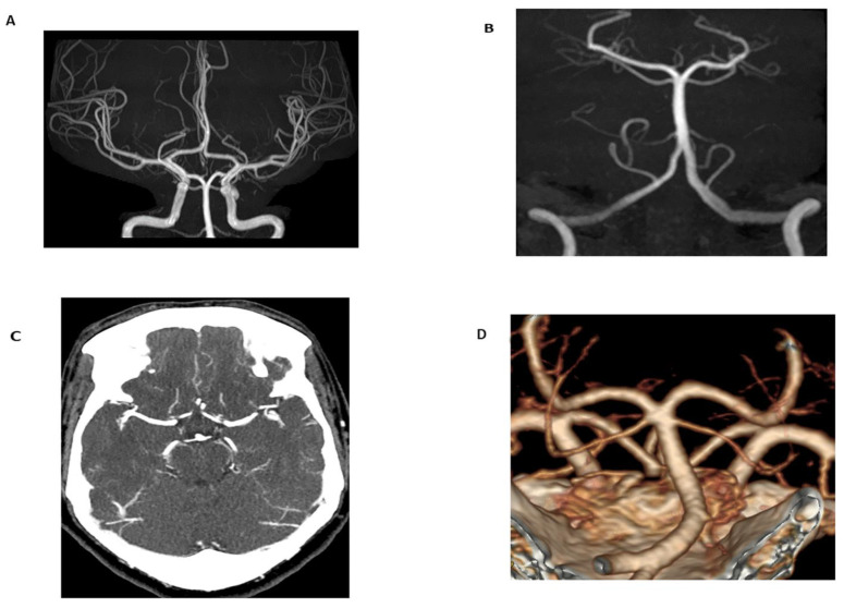
Figure 1.
Time-of-flight (TOF) maximum intensity projection (MIP) image of the Circle of Willis (A) and basilar artery (B) in a 24-year-old female obtained for migraines and dizziness. The magnetic resonance angiogram (MRA) demonstrated no flow-limiting stenosis or occlusion. Additionally, imaging was performed without radiation or intravenous contrast (technical specifications: FOV 200.00 mm, TR 25 ms, TE 3.5 ms, 3 T magnetic field strength). In comparison to TOF-MRA, axial CT angiogram (CTA) MIP images (C) and volume-rendered images (D) in a 60-year-old female obtained for evaluation of tinnitus demonstrated no flow-limiting stenosis or occlusion. 100 mL of Omnipaque 350 contrast was administered and the CTDIvol for the CTA of the head and neck was 59.6 mGy.

