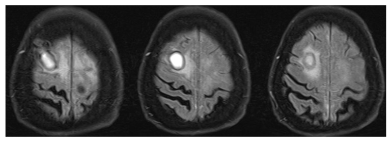
Figure 3.
Right frontal lesion associated with rheumatoid arthritis. Right frontal lesion on T2 FLAIR MRI sequence that was differentiated between non-typical abscess and neoplastic changes (lymphoma, melanoma metastasis). Brain biopsy revealed necrotizing granulomatous inflammation with pachymeningitis, findings compatible with CNS lesion due to rheumatoid arthritis. T2 FLAIR—T2-weighted-fluid-attenuated inversion recovery; MRI—magnetic resonance imaging; CNS—central nervous system.

