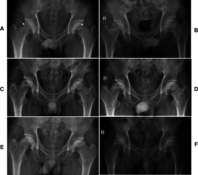
Figure 1.
The preoperative x-ray shows cystic and sclerotic changes in the bilateral femoral heads (white arrow) (A). A series of postoperative x-ray (B, immediate), (C, 3 months), (D, 6 months), (E, 12 months), and (F, 24 months) show a gradual healing of the necrotic area at the bilateral femoral heads.

