Figure 4.
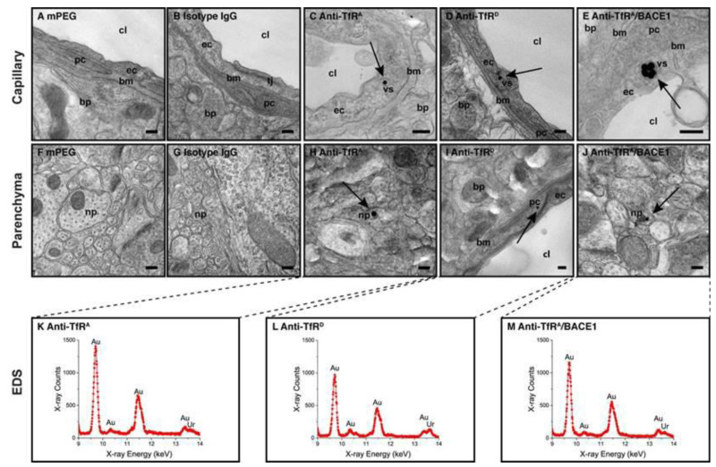
Detection of anti-transferrin receptor IgG conjugated gold-labeled nanoparticles (AuNPs) using transmission electron microscopy (TEM) in a normal adult mouse brain. Anti-transferrin receptor antibodies vary in affinity. (A,B) AuNPs are not detected in brain capillaries of mice in the mPEG (no IgG added) or isotype (non-immune) IgG groups. (C–E) In contrast, the AuNPs targeted with anti-transferrin receptor IgG are found in BECs (arrows). The AuNPs are detected in BECs confined to vesicular structures, suggesting receptor-mediated endocytosis as the uptake mechanism. (F,G) In brain parenchyma, AuNPs are not detected in mice in the mPEG or isotype (non-immune) IgG groups. (H–J) AuNPs are seen in brain parenchyma of mice treated with all transferrin receptor (TfR)-targeted variants, among which they are most easily detected in the anti-TfRA/BACE1 group (J). The sites for transport of the AuNPs may derive from transport across either BECs or post-capillary venules (see text body). All AuNPs detected in the brain parenchyma were analyzed using energy-dispersive X-ray spectroscopy (EDS) to validate the true presence of gold in the electron-dense points (K–M). Scale bars depict 200 nm. bp: brain parenchyma; bm: basement membrane; cl: capillary lumen; ec: endothelial cell; np: neural process; pc: pericyte; tj: tight junction; vs: vesicular structure. Modified from [54].

