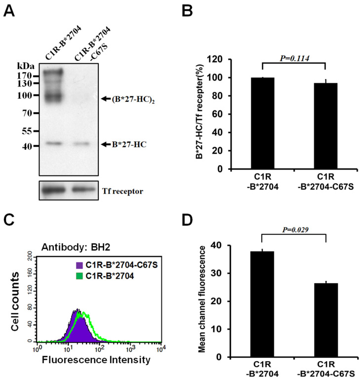
Figure 1.
Analyses of membrane-bound B*27-HC species of C1R-B*2704 or C1R-B*2704 C67S cells through Western blotting and flow cytometry. The membrane-bound proteins of C1R-B*2704 or C1R-B2704 C67S cells were labeled with biotin and isolated by pull-down using avidin beads. (A) Analysis of membrane-bound B*27-HC species via Western blotting. An aliquot (50 μg) of each protein extract was separated using non-reducing SDS-PAGE, followed by Western blotting using the BH2 monoclonal antibody. (B) The ratio of monomeric B*27-HC/transferrin receptor obtained from Figure 1A is plotted. Values (mean ± SD, n = 4) are averaged from four independent experiments. (C) The membrane-bound HLA-B*2704 C67S or HLA-B*2704 was pre-associated with BH2, stained with FITC-conjugated secondary antibody, and analyzed via flow cytometry. (D) The results obtained in Figure 1C are plotted. Values (mean ± SD, n = 4) are averaged from four independent experiments.

