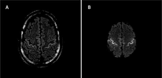
Figure 2:

MRI of the brain showing T2 Flair (A), and DWI (B), demonstrating restricted diffusion in the cortex at the pre- and post-central gyri bilaterally seen with rabies encephalitis evolution
MRI = Magnetic resonance imaging; FLAIR = Fluid-attenuated inversion recovery; DWI = Diffusion-weighted imaging
