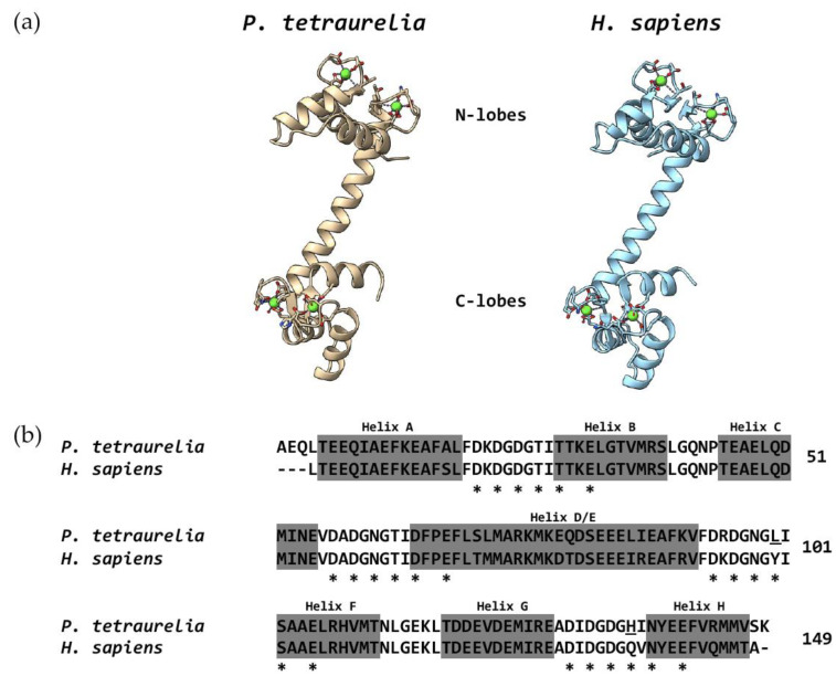
Figure 4.
Tertiary structure (a) and sequence comparison (b) between the CaM of Paramecium and humans. (a) Sequences were downloaded from the Protein Data Bank under the accession numbers 1OSA (P. tetraurelia) and 1CLL (Homo sapiens). Drawings using these sequences were made using Chimera X [152]. Green spheres represent Ca2+ ions. (b) The amino acid sequences in (a) were aligned using Chimera X. Note that these sequences are shorter than 149 amino acids since the crystals were obtained using non-full-length CaMs. Dashes indicate that the crystal structure lacked the amino acid at these positions. Stars indicate the amino acids that coordinate Ca2+ ions in humans according to [15]. Underlined amino acids mark those ones that are different in Paramecium at the Ca2+-coordinating sites. Grey-shadowed amino acids correspond to helices (see text). Numbers at the right indicate the amino acid position in the sequence of Paramecium.

