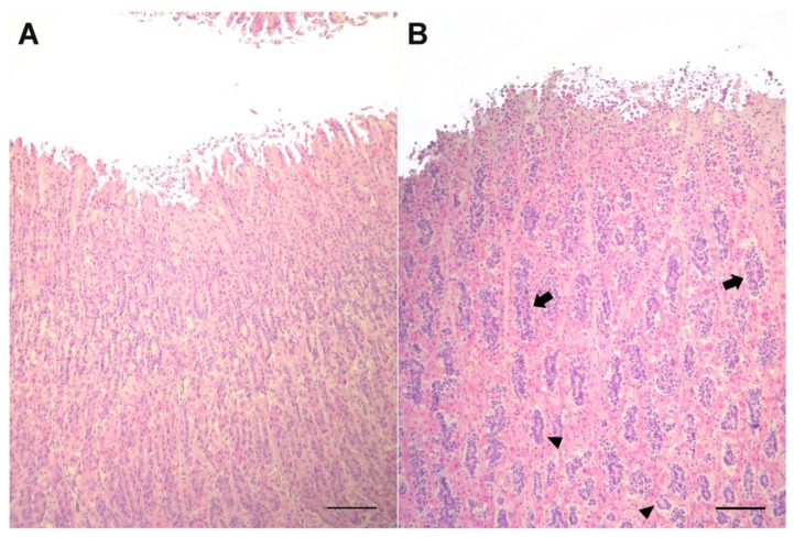
Figure 2.
Histological sections of the second stomach from bottlenose (A) and Risso’s (B) dolphins. At this level, mucosa shows peculiarities in the number and pattern of arrangement of the principal cells in Risso’s dolphin, with a higher concentration of principal cells, smaller and strongly stained by hematoxylin. Note the arrangement of these cells in cords (arrows) more superficially or in an acinar pattern at deeper positions (arrowheads) in Risso’s dolphin. H&E, bar = 500 µm.

