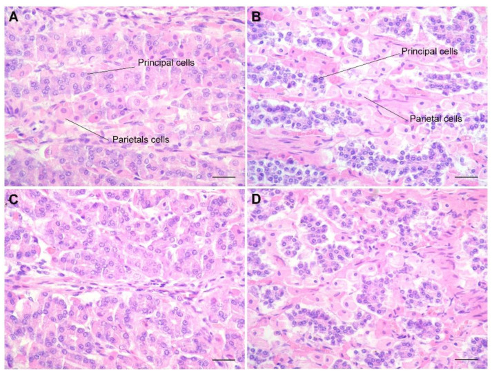
Figure 3.
Bottlenose (A,C) and Risso‘s (B,D) mucosa of the second stomach. At higher magnifications, the mucosa presents evident morphological differences. In Risso’s dolphin, the principal cells are considerably smaller and strongly stained by hematoxylin compared to the main cells of bottlenose dolphins. They are also located outside the parietal cells, not intercalated between them. At this magnification, the greater concentration is even more evident, but also the cordoniform (B) and acinar disposition (D) assumed by the principal cells, strongly basophilic, in the mucosa from Risso’s dolphin compared to bottlenose (A,C). H&E stain; bar = 250 µm.

