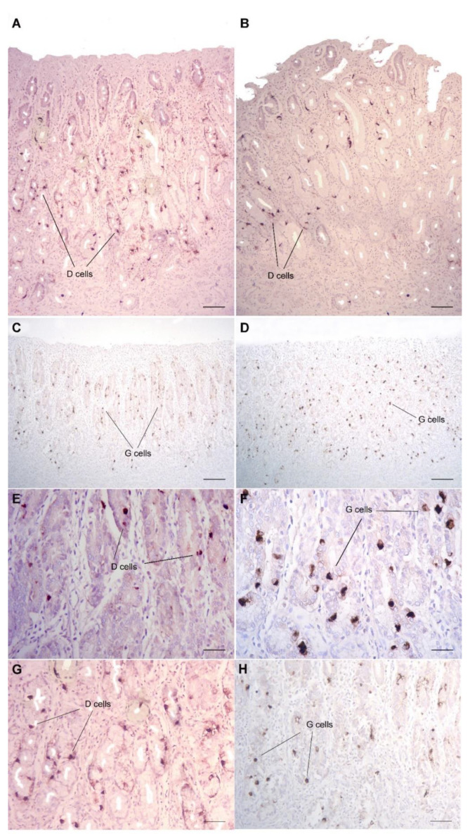
Figure 4.
Immunostaining of G and D cells in bottlenose (A,C,G,H) and Risso’s gastric mucosa (B,D–F) of the second stomach. Although presenting a similar distribution pattern, Risso’s D cells were few and scattered in the mucosa (B,E), while its G cells were very abundant and diffusely expressed in the same mucosal areas (D,F). In contrast, in bottlenose dolphin, G and D cells were equally represented and alternated at the level of the mucosa (A,C,G,H). Anti-G-cell and anti-D-cell immunostaining, Harris’s hematoxylin counterstain, bar = 500µm (A,B), 600 µm (C,D) and 150 µm (E–H).

