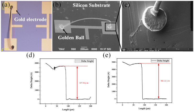
Figure 3.
Analysis of interface and electrodes after ball bonding. (a) Photo of electrodes and integrated bonding optical microscopy. (b) Scanning Electron Microscope Images of Electrodes and Global Bonding. (c) Interface Diagram of Spherical Welding under High Power Scanning Electron Microscope. (d) Measuring 50/300 nm thick Cr/Au electrodes with a step instrument. (e) Measuring 50/400 nm thick Cr/Au electrodes with a step instrument.

