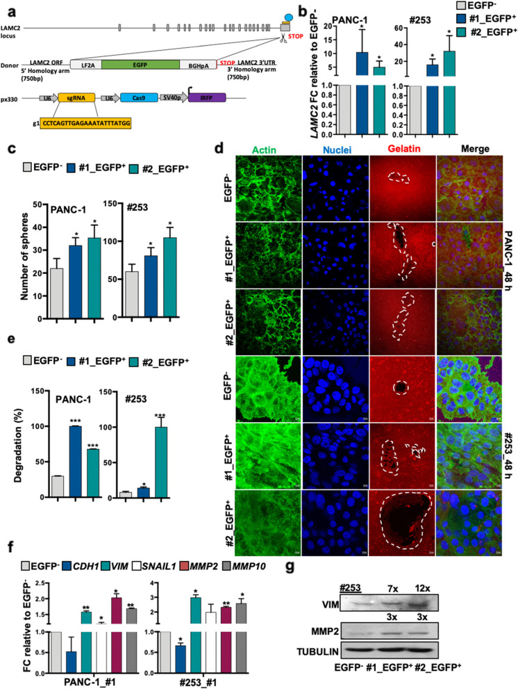
Fig. 4.
Generation of LAMC2-EGFP knock-in human pancreatic cancer cells. a Design of LAMC2‐EGFP donor and CRISPR/Cas9 sgRNA vectors. Blue circle represents the CRISPR/Cas9 protein complex and the yellow box underneath illustrates the guide RNA. b qPCR analysis for LAMC2 gene expression in EGFP+ and EGFP− cells. Data are normalized to GAPDH and are presented as fold change in gene expression relative to the EGFP− counterpart. c Sphere formation capacity for EGFP+ versus EGFP− cells. d Representative images of gelatin degradation for EGFP+ versus EGFP− cells. Nuclei were stained with Hoechst 33342 (blue), green represents actin (Alexa Fluor™ 488 Phalloidin) and red illustrates gelatin (Rodhamine). The white dashed line circles indicates the areas of degradation. e Invasive potential of sh empty versus LAMC2 knockdown cells. f qPCR analysis for EMT and MMP2 and MMP10 gene expression for EGFP+ versus EGFP− cells. Data are normalized to GAPDH and are presented as fold change in gene expression relative to the EGFP− cells. g Western blot analysis of VIM and MMP2 in EGFP+ and EGFP− cells. Parallel Tubulin immunoblotting was performed. *p < 0.05, **p < 0.005, ***p < 0.0005. n Statistical significance was assessed by Student's t-test

