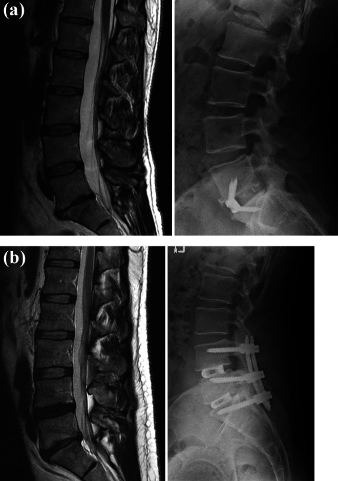
Figure 2.

Illustrative cases: pre-operative magnetic resonance imaging (MRI, T2 sagittal) and 12-month post-operative xrays of (a) a 36-year old female who presented with back pain and degenerative disc disease at L5-S1, and (b) a 41-year old male who presented with back pain and degenerative disc disease at L4-5 and L5-S1. Both patients experienced improvement of their back pain that exceeded the Minimum Clinically Important Difference.
