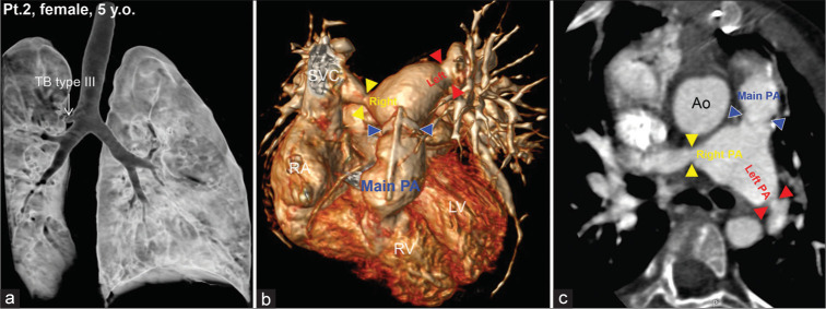
Figure 3:
Chest computed tomography images of a 5-year-old girl with a tracheal bronchus on the right side, at the site of the carina (type III) (a). There was stenosis at the main (blue arrow heads), right (yellow arrow heads), and left (red arrow heads) pulmonary arteries (b and c). right atrium, right ventricle, left ventricle, Ao, aorta, pulmonary artery, and superior vena cava.

