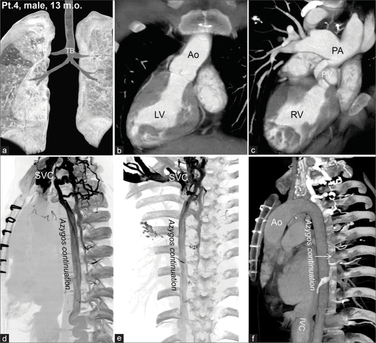
Figure 5:
Computed tomography images of a 13-month-old boy with bilateral tracheal bronchus and bilateral main bronchial stenosis (a). This patient had a right-sided heart (b), abnormalities in the pulmonary valve (PV), main pulmonary artery stenosis, and dilatation of the right and left pulmonary arteries (c). An azygos continuation (d-f) was observed due to interrupted inferior vena cava (IVC), commonly associated with the left isomerism. PV, (pulmonary valve)

