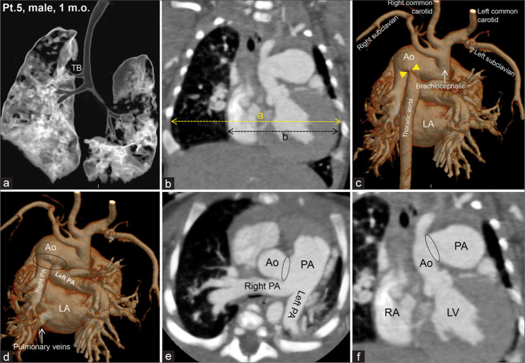
Figure 6:
Computed tomography images of a 1-month-old boy with a tracheal bronchus on the right side (Type II) (a). The patient had an enlarged heart (b), in which the cardiac–thoracic index = the maximum transverse diameter of the heart/the maximum internal diameter of the thorax = b/a >0.6 (for newborns). A brachiocephalic trunk arising from the aortic arch was observed, from which the bilateral subclavian and common carotid arteries arose. The thoracic aorta was severely narrow (yellow arrow heads) at the origin, emerging from the aortic arch (c). The root of the pulmonary artery has a large opening (oval circle) to the aorta (d-f).

