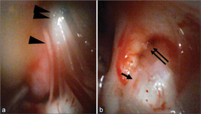
Figure 2:

Observations of an angioscope inserted into the cerebellopontine angle Panel (a) depicts the use of the angioscope to observe the glossopharyngeal (arrow head) and vagus nerves (double arrow heads) in the cerebellopontine angle. Panel (b) indicates the use of the angioscope to observe the trigeminal nerve (arrow) and Meckel’s cavity (double arrows).
