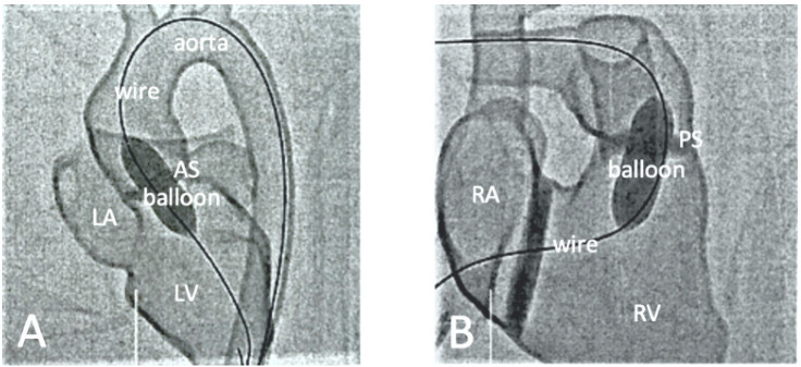
Figure 6.
Fluoroscopic documentation of balloon dilatation of valvular stenosis with the 3D printed heart model. (A): Balloon dilatation of a valvular aortic stenosis. The inflated balloon is positioned at the level of the aortic valve. The long guidewire is inserted via the descending aorta with its tip lying in the LV. (B): Balloon dilatation of a valvular pulmonary stenosis. The inflated balloon is positioned at the level of the pulmonary valve. The long guide wire is inserted via the inferior vena cava through the RA into the RV, with its tip lying in the right pulmonary artery. AS-aortic stenosis, PS-pulmonary stenosis, LA-left atrium, LV-left ventricle, RA-right atrium, RV-right ventricle. Reprinted with permission under the open access from Brunner et al. [65].

