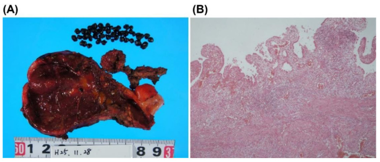
Figure 3.
Macroscopic and microscopic findings of the gallbladder. (A), Macroscopic finding of the resected gallbladder. More than 30 black stones of 2–3 mm in diameter were found in the gallbladder. The base of the gallbladder was partially melted due to necrotizing cholecystitis and abscess. (B), Microscopic findings revealed erosion, fibrosis, and moderate inflammatory cell infiltration with neutrophils, but no malignant cells.

