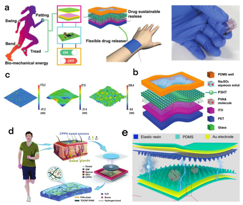
Figure 12.
(a) The TENG collects biomechanical energy to provide power for the FDRD. (b) Structure of the FDRD. (c) The surface morphology of the ITO layer, the PVA molecular layer, and the P3HT layer was recorded with an atomic force microscope (AFM) [87]. Liu et al. (2020), John Wiley and Sons Inc. (d) Cellulosic conductive hydrogel for self-powered sweat measurement, CPPH electrode material, microstructure diagram, and CPPH sweat sensor module structural diagram. During body movement, the sweat sensor monitors the concentration of sweat ions in real time. The CPP sweat sensor is placed on the skin’s sweat glands to detect and quantify Na+, K+, and Ca2+ [88]. Qin et al. (2022), John Wiley and Sons Inc. (e) Schematic structure of the BSRW-TENG [89]. Li et al. (2022), Elsevier.

