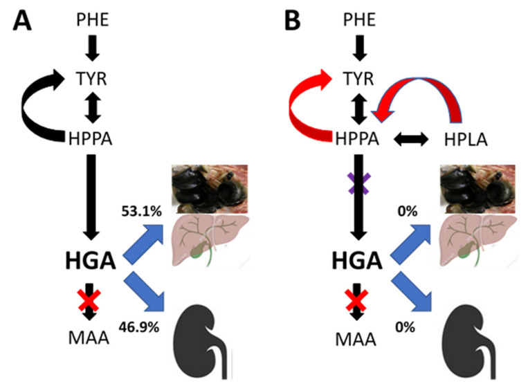
Figure 4.
Diagrammatic representation showing that HGA accumulates in AKU due to a deficiency of homogentisate dioxygenase (denoted by the red X), with a measured spill-over into urine as well as predicted biliary excretion and accumulation in the body as pigment. In AKU (Panel (A)), circulating HGA is in low concentration relative to urinary excretion and therefore the urinary excretion is dominant. However, data during nitisinone (reflecting 4-hydroxyphenylpyruvate dioxygenase inhibition denoted by the purple) show that 53.1% of TYR flows into the non-urinary pathways including biliary and pigment pathway and only 46.9% is excreted in the urine, following 10 mg nitisinone daily for 4 weeks. Panel (B) shows the complete inhibition of HGA urinary and biliary excretion as well as HGA deposition and the suggested mechanism for metabolite generation during nitisinone treatment. (PA: phenylalanine; TYR: tyrosine; HPPA: 4-hydroxyphenylpyruvate; HPLA: 4-hydroxyphenyllactate; HGA: homogentisic acid; MAA: maleylacetoacetic acid) (Image of pigmented elbow joint denotes the total body ochronotic process; that of the liver denotes the biliary excretion, while the kidney image denotes urinary HGA excretion.)

