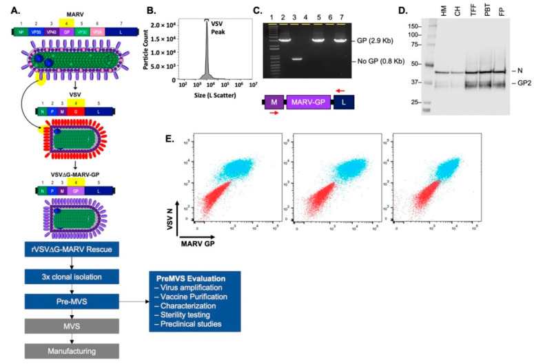
Figure 1.
Generation and characterization of the rVSV∆G-MARV-GP vaccine for use in humans. (A) The schematic summarizes the rVSV∆G-MARV-GP chimeric virus design and critical work stages leading to cGMP MVS production and vaccine manufacturing. Following the regeneration of a new recombinant rVSV∆G-MARV-GP virus, 3 rounds of virus plaque isolation were performed prior to the generation and evaluation of a pre-MVS. (B) Nanoflow cytometry using an Apogee instrument was performed [51] with rVSV∆G-MARV-GP virions purified using tangential flow filtration (TFF). The graph shows particle counts (Y axis) and large-angle light scatter (X-axis). Total particles for the vaccine candidate were 6.7 × 10¹⁰ particles/mL, VSV virus peak (indicated) corresponded to 73.2% of the total particles. (C) GP gene integrity was evaluated by RT-PCR using primers specific for the VSV M and L genes that flank the GP insert. Analysis of PCR products by agarose gel electrophoresis showed that the expected 2.9 Kb product was amplified and that no larger or small products were detectable. Samples analyzed on the gel include: (1) 1 Kb ladder; (2) positive control VSV-MARV genomic plasmid template; (3) a negative control VSV genomic plasmid DNA (pVSV-G5) in which the G gene was moved to the 5′ terminus of the genome and no transcription unit is present between M and L [52]; (4) A negative control containing no template nucleic acid in RT-PCR reaction; (5) Medium harvest containing rVSV∆G-MARV-GP released by infected cells; (6) Empty well; (7) Purified rVSV∆G-MARV-GP. (D) Western blot analysis conducted using samples from various stages of virus purification. Blot probed with anti-GP rabbit polyclonal antisera that bound to MARV GP2 and rabbit poly-clonal anti-VSV-N. Analyzed samples included: Harvested medium (HM); clarified harvest (CH); product concentrated by TFF (TFF); product post-benzonase treatment (PBT); and final product (FP). (E) Flow cytometry was performed using Vero cells infected with purified rVSVΔG-MARV-GP vaccine material. Overlay dot plots displaying co-expression of MARV GP (x-axis) and VSV-N (y-axis) on uninfected VERO cells as controls (red population) and VERO cells 40 h post infection (MOI 0.001) with rVSV∆G-MARV-GP (blue population). GP was detected with three different anti-GP Abs (Pan-Filovirus anti-GP mAb (left panel), murine mAb 5C1 (middle panel), or rabbit anti-GP pAb (right panel)), and intracellular VSV-N was detected with murine mAb 10G4.

