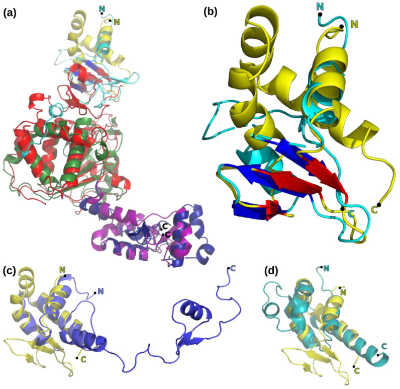
Figure 5.
Structures related to D5323–785. N and C-terminal extremities of the fragments are labeled. (a) Flexible structural alignment using the FATCAT server of the NrS-1 helicase domain (pdb entry 6k9c) with D5323–785 (domains colored as in Figure 1, the 3 β-strands in the collar domain are colored in red). Cyan, green, and blue are used respectively to color the NrS-1 helicase with the 3 β-strands shown in blue. The FATCAT alignment creates chain breaks for the molecule submitted to the alignment. (b) Zoom on the superposition of the collar domains of panel (a). (c) Similarity between the collar domain of D5 (yellow) and a part of the ribosomal protein S17 (pdb entry 6zxh, blue). (d) Similarity between the collar domains of D5 (yellow) and papillomavirus E1 helicase (pdb entry 2gxa, turquoise). Illustration made with PyMol.

