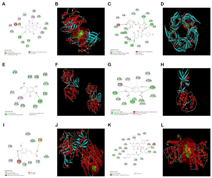
Figure 14.
Docking results of six compounds with the highest matching target proteins: EA-GSK3β (A,B), KA-α7nAchR (C,D), GA-CDK5 (E,F), VA-CDK5 (G,H), FA-γsecretase (I,J), TI-PDE4A (K,L). 2D molecular docking diagrams are represented by (A) Ellagic acid, (C) Kaempferol-3-o-rutin glycoside, (E) Gallic acid, (G) Vanillic acid, (I) Ferulic acid, and (K) Tiliroside; and 3D molecular docking diagram are represented by (B) Ellagic acid, (D) Kaempferol-3-o-rutin glycoside, (F) Gallic acid, (H) Vanillic acid, (J) Ferulic acid, and (L) Tiliroside, respectively.

