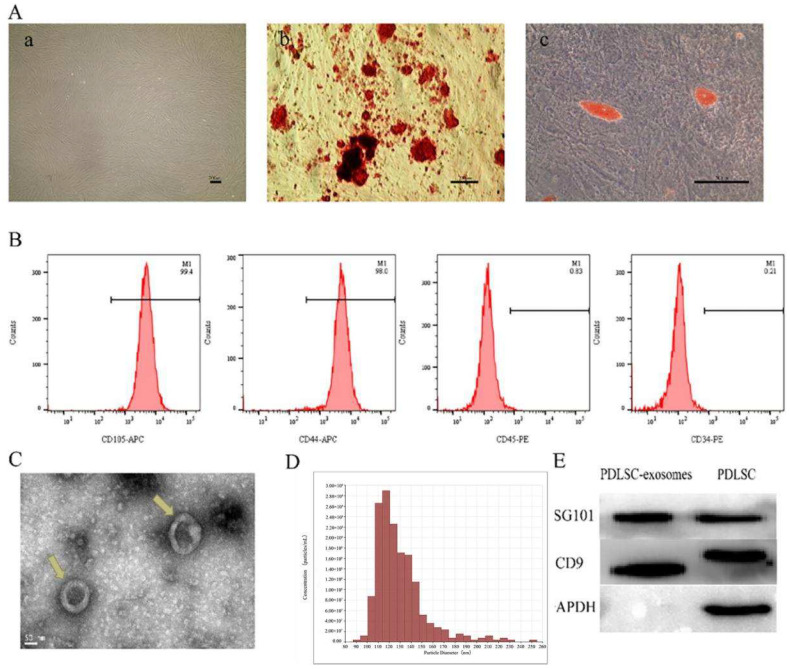
Figure 1.
Characterization of PDLSCs and PDLSCs-derived exosomes. (A) Characterization of PDLSCs (a) Representative images of PDLSCs. Spindle shape with vortex distribution (b) Representative images of osteogenesis of PDLSCs stained with Alizarin red staining. A large number of red mineralized nodules were observed. (c) Representative images of adipogenesis of PDLSCs stained with Oil Red O. Red lipid droplets were formed. (B) Flow cytometric analysis of surface markers CD105, CD44, CD45, CD34 in PDLSCs. CD105 and CD44 are highly expressed, while CD45 and CD34 are low expressed. (C) Exosomes morphology of a cup-shape or circle observed by TEM (indicated by the yellow arrowheads). (D) Particle size distribution of exosomes concentrated between 90 and 150 nm detected by TRPS. (E) Western blot analysis of the exosomal markers TSG101 and CD9.

