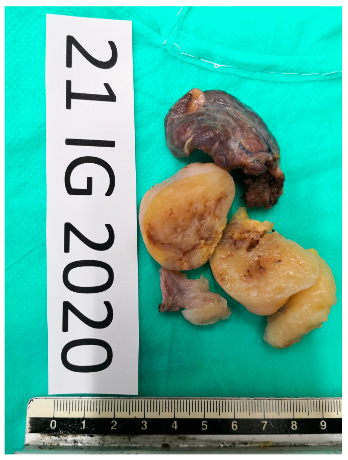
Figure 5.
Definitive pathology showing mesenchymal proliferation, spindle-cells type, with both dense and loose areas with thickened walls vases at hematoxylin and eosin staining. In dense areas (Antoni A), the so-called “Verocay bodies” were evident, while in the loosest areas (Antoni B), they appeared evident diffuse nuclear atypia.

