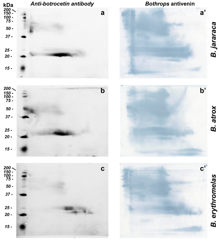
Figure 7.
Western blotting of B. jararaca (a,a’), B. atrox (b,b’) and B. erythromelas (c,c’). The venom proteins were subjected to BN/SDS-PAGE. In the left column (a–c), nitrocellulose membranes were incubated with the anti-botrocetin polyclonal antibody, the anti-rabbit IgG conjugated with AlexaFluor 647, and then analyzed in a fluorescence detecting system. Thereafter, each membrane was incubated with Bothrops antivenin and developed with DAB (right column, (a’–c’)).

