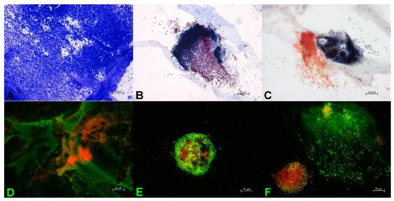
Figure 2.
Microscopic evidence of the mature 48 h multispecies 3D biofilm with S. aureus, E. coli, and A. baumannii. (A) Staining with crystal violet confirms dense microbial population. Gram staining reveals a close proximity of clustered populations of the Gram-positive S. aureus (dark purple) and the Gram-negative E. coli (reddish purple) (B), or the Gram-negative A. baumannii (reddish) (C). Staining with TOTO-1/SYTO 60 (D–F) shows on the one hand the clustered growth of the microorganisms with a very vital, red center (D–F). On the other hand, the external DNA of the extracellular matrix is visible as long green DNA strands (D,F). In (F), the close proximity of a S. aureus cluster (left) and E. coli cluster (right, surrounded by large amounts of external DNA) is visible.

