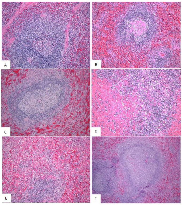
Figure 9.
Representative images of microscopic findings: Spleen. Microscopic findings at necropsy in cynomolgus macaques intramuscularly exposed to SUDV Gulu. Spleen. (A) Day 21 control, No. 478. Essentially normal tissue. 4x; (B) Day 2 PE scheduled necropsy, No. 491. Follicles contain increased hyaline material, consistent with spontaneous change. 20x. (C) Day 5 PE scheduled euthanasia, No. 493. There is moderate lymphoid depletion with deposition of fibrin within the perifollicular marginal sinus. 20x; (D) Day 7 PE scheduled necropsy, No. 477. There is moderate lymphoid depletion with diffuse fibrin deposition. 20x; (E) Day 9 PE scheduled necropsy, No. 476. There is lymphoid depletion with lymphocytolysis and marginal sinus fibrin deposition that extends into the red pulp. 20x; (F) Day 21 PE SUDV-exposed survivor, No. 487. Follicles are enlarged with prominent germinal centers (follicular hyperplasia). 10x.

