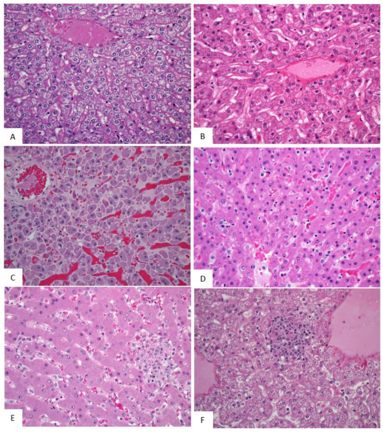
Figure 10.
Representative images of microscopic findings: Liver. Microscopic findings at necropsy in cynomolgus macaques intramuscularly exposed to SUDV Gulu. Liver. (A) Day 21 control, No. 478. Essentially normal tissue. 40x; (B) Day 2 PE scheduled euthanasia, No. 491. Essentially normal tissue. 40x; (C) Day 5 PE scheduled euthanasia, No. 493. There is single cell hepatocellular degeneration, necrosis and vacuolation. 40x; (D) Day 7 PE, No. 477. There is rare necrosis of individual hepatocytes with inflammation and sinusoidal fibrin. 40x. (E) Day 9 PE scheduled euthanasia, No. 476. There is single cell hepatocellular necrosis with inflammation and fibrin. 40x; (F) Day 21 PE SUDV survivor, No. 487. There is multifocal mononuclear cell inflammation. 40x.

