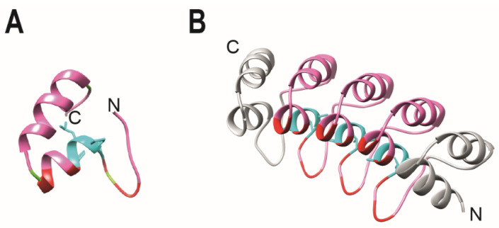
Figure 3.
Structure of a monoDARPin domain. (A) Library module. The library module is colored in pink, with the conserved TPLH residues in cyan. Residues then can be randomized are indicated in red. (B) Homology model of monovalent DARPin R2 generated by SWISS MODEL (SIB) using the consensus designed ankyrin repeat domain PDB ID 2xee and the sequence of mono DARPin R2 [39]. The N-cap and C-cap are colored in grey.

