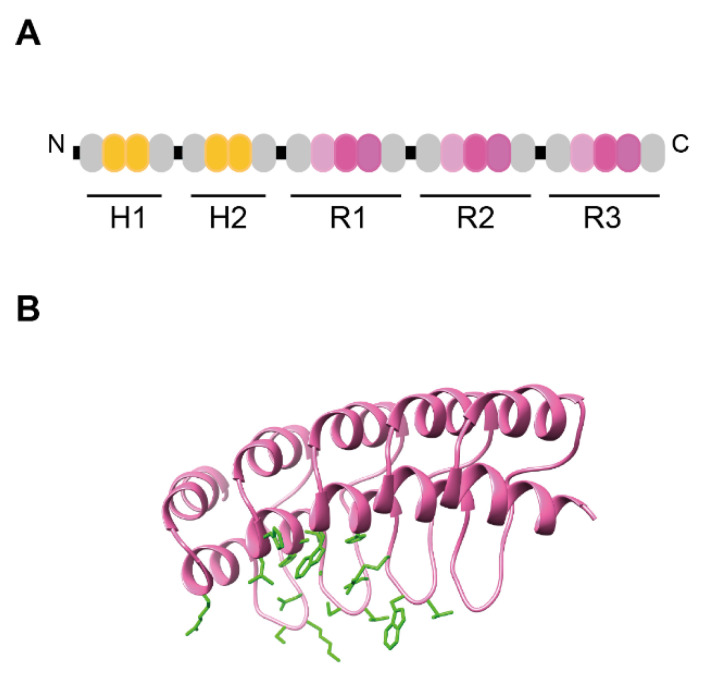
Figure 5.
Schematic representation of ensovibep and structure of monoDARPin R2. (A) Schematic representation of ensovibep. The protein comprises two HSA-binding DARPin domains for in vivo half-life extension and three DARPin domains that bind the RBD of the SARS-CoV-2 spike trimer. The HSA-binding DARPins contain two internal repeats (indicated in yellow) and the RBD binding DARPins three internal repeats (indicated in pink). The N-Cap and C-Cap repeats are represented in grey. The linker sequences are colored in black. (B) Binding surface of monovalent DARPin R2. Homology model of monovalent DARPin R2 generated by SWISS MODEL (SIB) using the consensus designed ankyrin repeat domain PDB ID 2xee and the sequence of DARPin R2 [39]. The library modules are colored in pink. The side chains of the residues interacting with the RBD of the SARS-CoV-2 spike trimer, as defined by cryo-EM analysis are highlighted in green.

