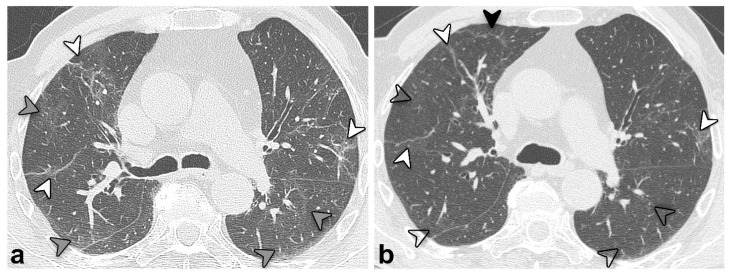
Figure 3.
Unenhanced chest CT scan axial images of the same patient in Figure 2 (group 3) at 3 (a) and 12 months (b) after discharge. An overall improvement is evident, with previous GGO (grey arrowheads, (a)) almost resolved but showing a “melting sugar” appearance on the latter (empty arrowheads, (b)). Reticular opacities (white arrowheads, (a,b)) also improved but remained evident; the black arrowhead (b) highlights a new onset of tiny peripheral reticular opacification.

