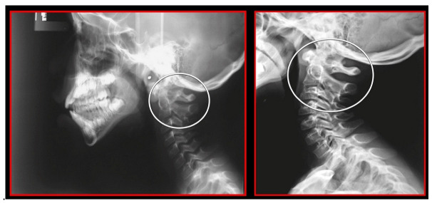
Figure 9.
Rotation of the atlas on a lateral cephalometric radiograph. Left: Pre-treatment. Double ring of the atlas showing its rotation. Decreased functional spaces. Right: After orthopedic manual therapy. Only one posterior arch of the atlas showing the normalization of its rotation. Normalized functional spaces.

