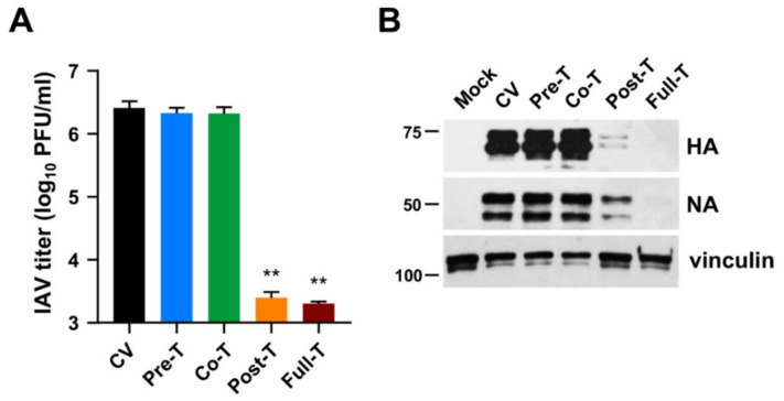
Figure 4.
MEDS433 targets a post-entry phase of the IAV replicative cycle. MDCK cell monolayers were infected with IAV at an MOI of 0.1 and, where indicated, treated with 0.5 μΜ MEDS433 1 h prior to the infection (from −2 to −1 h, Pre-T), or during the infection (from −1 to 0 h, Co-T), or after infection (from 0 to 48 h p.i., Post-T;), or from −2 to 48 h p.i. (Full-T). Mock-infected cells (Mock) and control IAV-infected cells (CV) were exposed to DMSO only. (A) Cell supernatants harvested at 48 h p.i. were titrated by the plaque assay, and viral plaques were microscopically counted and plotted as PFU/mL. The data shown are the means ± SD of two independent experiments performed in triplicate and analyzed by a one-way ANOVA followed by Dunnett’s multiple comparison test. ** (p < 0.001) compared to the calibrator sample (CV). (B) Total cell extracts were prepared at 48 h p.i., fractionated by 8% SDS-PAGE, and analyzed by immunoblotting with anti-IAV HA and anti-IAV NA pAbs. Vinculin immunodetection was used as a control for protein loading. Molecular weight markers are shown next to the left side of each panel.

