Figure 2.
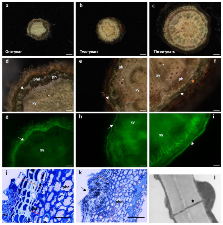
Cork develops as early as one-year stems in Quercus suber. (a–i) Cross-sectional stereomicroscopic images of one-, two- and three-year-old stems. (Scale bars: (a,b) 100 µm, c 500 µm). (d–f) Details of the xylem with empty tracheids (*). Moving outwardly, one finds the phloem (dark layer) surrounded by the periderm comprising the phelloderm (orange arrow -phd), a thin layer of phellogenic cells (white arrowhead) and several rows of suber cells. The most external layer is the remaining epidermis. (Scale bars: (d,e) 100 µm, f 500 µm). (g–i) Cork cells exhibit autofluorescence when excited with UV light [18]. The stems observed in d-f were excited with UV using a stereoscope filter for “Lumar01” (BP 365/12; LP397) after which, the suberin layer is clearly distinguishable (white arrow). In older stems, as xylem cells mature, autofluorescence can be observed since lignin also possesses some autofluorescence properties under UV light. (j,k) Light microscopy of one-year stem cross sections stained with toluidine blue. (Scale bars: (g,h) 100 µm, i 500 µm). (j) Some remaining epidermis (ep) is still present and right below, six to seven cell cork layers with cells displaying a hyaline cell wall appearance and filled with electrodense material (phenolic compounds) (orange line) can be observed. Inwardly and adjacent to suberin cells, is the phellogen composed of only one to two cells (red line). Cells are filled with cytoplasm and cell walls do not present a secondary thickening. Right below is the phelloderm with the characteristic round-shaped parenchymatous cells. (Scale bar: 10 µm). (k) Cross section showing a lenticel forming in a one-year stem (black arrow). The disorganized division of the meristematic cells inside the lenticel structure is starting to push the suberin layer outwards leading to a complete rupture of this layer to form an aperture that allows gas exchanges. (Scale bar: 40 µm). (l) Transmission electron microscopy image of amadia cork cell wall clearly showing a plasmodesmata (black arrow) crossing both suberized cell walls. It is possible to observe cytoplasmic deposition in the inner part of the cell, with thickening appearing on both sides of the plasmodesmata channel (amp. 20,000×). ep—epidermis; ph—phloem; phd—phelloderm; phl—phellogen; s—suberin; xy—xylem.

