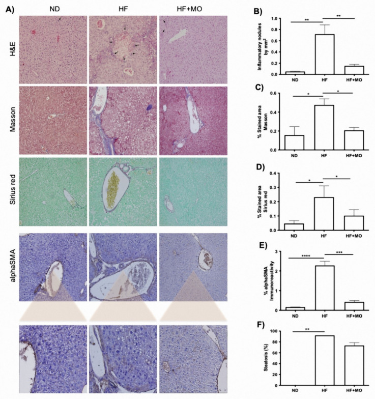
Figure 3.
Liver histology showed improved parameters after Mo supplementation. (A) Representative microphotographs (40X) of liver tissue stained with H&E, Masson, Sirius Red and after IHQ against αSMA. (B) Quantity of inflammatory nodules is reduced in Mo animals compared to HF group (p < 0.01). (C,D) showed a decrease in ECM and collagens after Mo supplementation (p < 0.05). (E) Positivity to αSMA is increased only in HF animals (p < 0.001). (F) steatosis tent to be reduced in Mo animals. Data are expressed as mean ± SE. * p < 0.05; ** p < 0.01; *** p < 0.001; **** p < 0.0001.

