Figure 3.
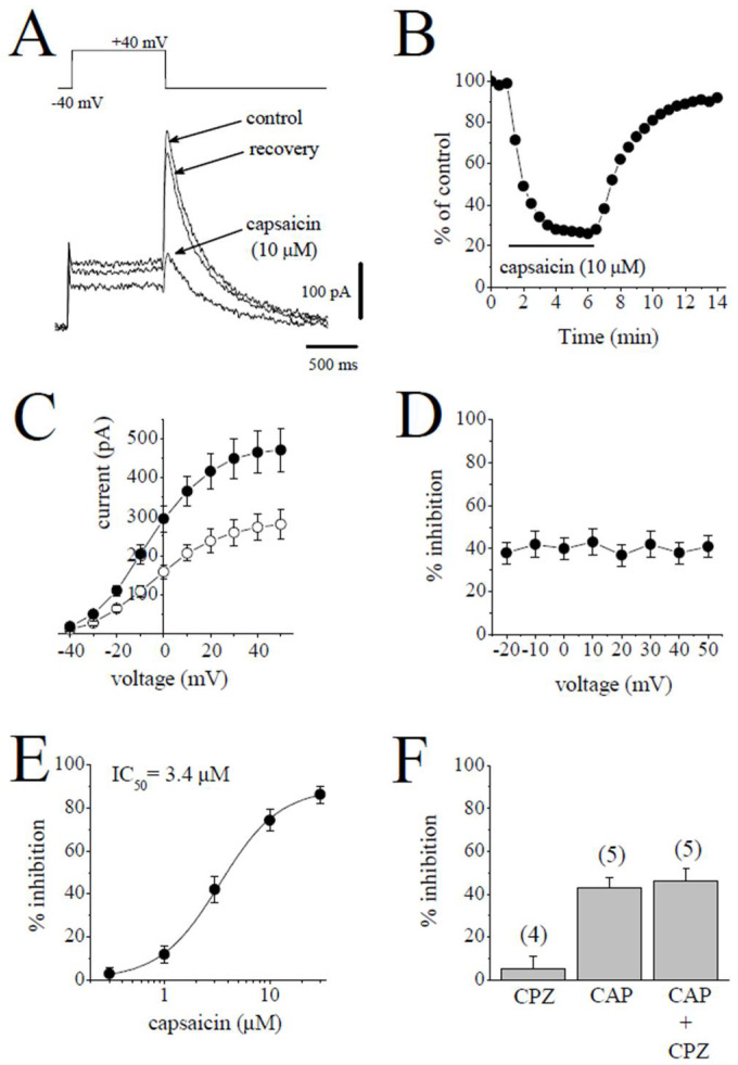
Capsaicin suppresses the IKr tail current. (A). Superimposed current traces in control, after 6 min exposure to 10 μM capsaicin and recovery. IKr tails were measured as a time-dependent component of the tail current activated in response to membrane repolarization. The pulse protocol to activate IKr is presented as an inset. (B) Time course of the effect of capsaicin effect on the maximal amplitudes of IKr and washout presented as % of control calculated from the mean of three control currents (n = 5). (C) Current–voltage relationships of IKr in absence (filled circles) and presence (open circles) of 3 µM capsaicin are shown (n = 6). (D) The relationship between test potential and the capsaicin (3 µM) inhibition of IKr (n = 6; p > 0.05, ANOVA). (E) Concentration-inhibition curve for capsaicin inhibition of IKr tails (n = 4–8). (F) The effect of capsazepine (3 µM) alone and the extent of capsaicin (3 µM) inhibition of IKr in the absence and presence of 3 µM capsazepine. The number of cells tested for each group was presented on top of each bar (p > 0.05; t-test).

