
Figure 1.
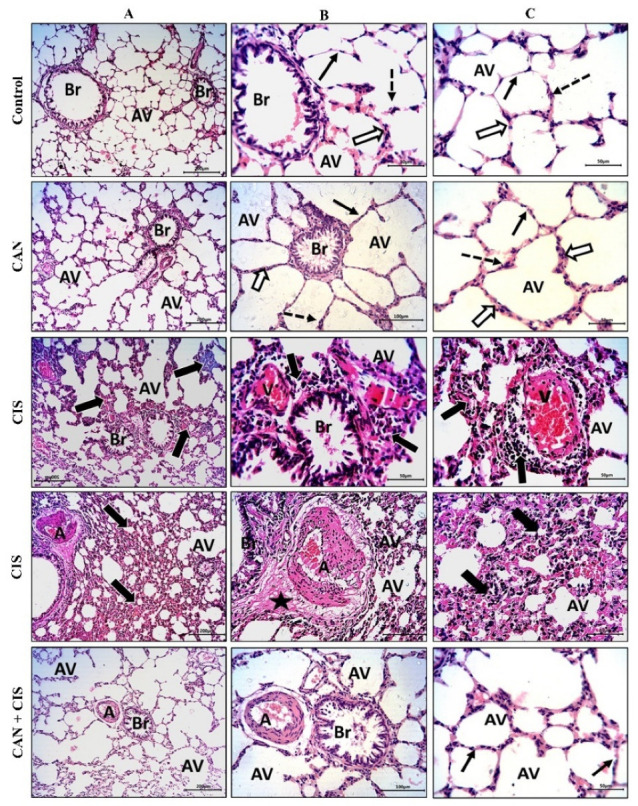
CAN prevented CIS-induced lung tissue injury. Photomicrographs of H&E-stained sections showing alveoli (AV) and bronchioles (Br) at ×100 (A), and at ×200 magnification (B,C). Control and CAN-treated rats showed normal alveoli lined with a thin type I alveolar epithelium (black arrows) with occasionally prominent rounded nuclei of type II cells (dotted arrows), thin interalveolar septa with no inflammatory cells (white arrows), and bronchioles (Br) with intact epithelium and lumen free of cell debris or secretions (Figure 1A–C). CIS-administered rats exhibited a decrease in the number of potent alveoli (Av), thick interalveolar septa with inflammatory cells (thick black arrows), congested veins (V) with perivascular mononuclear infiltrate (thick black arrows), thickened and deformed arteries (A), and perivascular edema (star). CAN-treated CIS-administered rats showed a significant improvement in the alveoli (AV), bronchioles (Br), and interalveolar septa (thin black arrows).