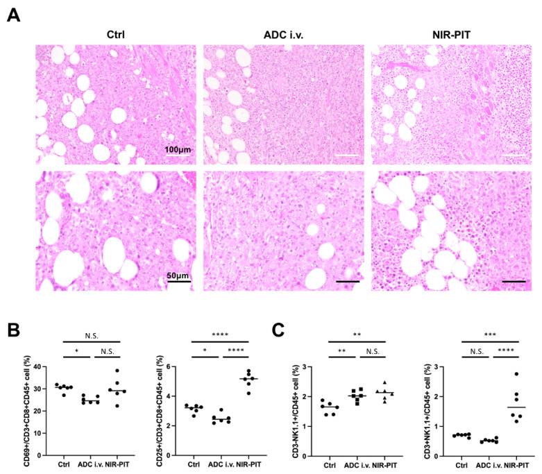
Figure 4.
Pathological analysis of EL4-luc tumor after GD2-targeted NIR-PIT. (A) Hematoxylin and eosin staining of tumors resected one day after NIR-PIT (White scale bars, 100 μm; black scale bars, 50 μm). (B,C) Immune cell response after GD2-targeted NIR-PIT was analyzed by flow cytometry 1 day after NIR-PIT. (B) Activation marker expression in CD8+ killer T cells in the regional lymph nodes. (n = 6; one-way ANOVA followed by Tukey’s test; * p < 0.05, **** p < 0.0001; N.S., not significant) (C) Number of NK and NKT cells in the regional lymph nodes. (n = 6; one-way ANOVA followed by Tukey’s test; ** p < 0.01, *** p < 0.001, **** p < 0.0001; N.S., not significant).

