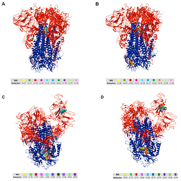
Figure 4.
Molecular simulation of tripeptides interacting with HCoV-OC43 and SARS-CoV-2 spike protein obtained by HPEPDOCK server: (A) TLH/HCoV-OC43 spike protein; (B) VFI/HCoV-OC43 spike protein; (C) TLH/SARS-CoV-2 spike protein; (D) VFI/SARS-CoV-2 spike protein. The red color indicates the S1 subunit and the blue color the S2 subunit of the spike protein. The different color code of peptides, represented as balls, refers to the different binding free energy.

