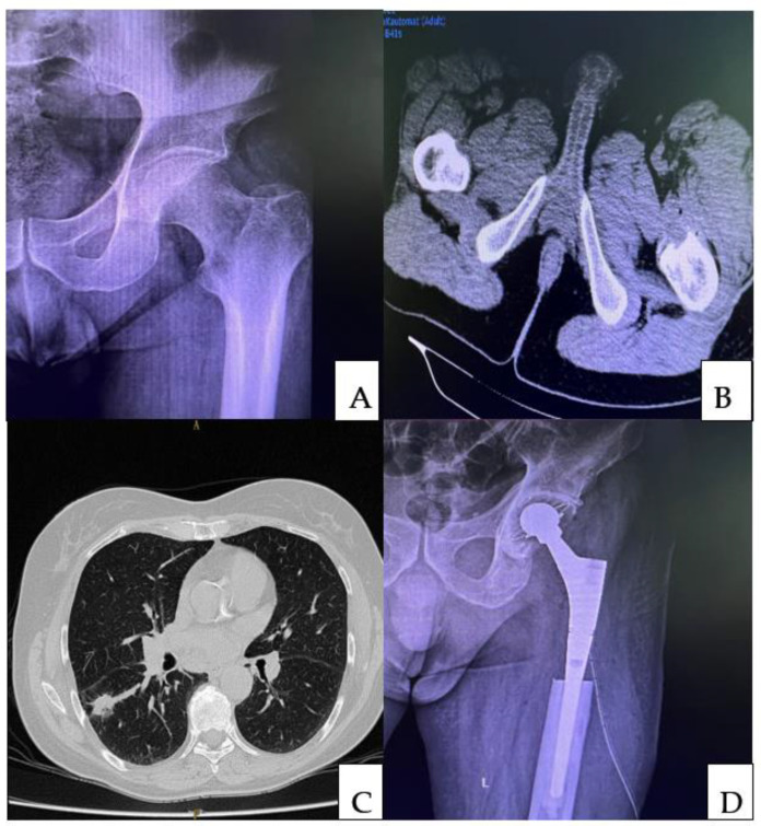
Figure 2.
Pre-operative X-ray (A) of a 49-year-old male complaining of pain in the left hip that started three months earlier. An area of osteolysis at the level of the lesser trochanter was detected on the radiograph of the left hip. A computed tomographic (CT) scan of the pelvis (B) was performed, which revealed an osteolytic tumor at the level of the lesser trochanter. Following the biopsy, a diagnosis of bone metastasis secondary to a lung carcinoma was made. A chest CT scan revealed a spiculated, iodophilic nodule with retraction of the overlying pleura at the level of the apical segment of the right lower lobe (S6—Fowler) (C). The preoperative staging was a pulmonary tumor with single bone metastasis. Following multidisciplinary consensus, the patient was subjected to the radical resection of the pulmonary tumor followed by reconstruction of the left proximal femur (D).

