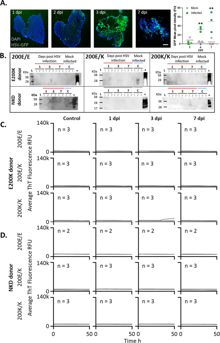
Fig 3. Acute HSV1 infection does not cause production of misfolded PrP or prion seeding activity accumulation.
Organoids were infected with a 0.00001 MOI of McKrae HSV1. A. Emergence of HSV1-GFP over 1–7 dpi demonstrating active viral replication and changing organoid morphology by 7 dpi. Scale bar = 500 μm. Quantification of GFP fluorescence in organoids over time indicating production of virus is shown right. Individual points show single organoids (n = 3) and horizontal lines show the average intensity. **p<0.01 from the background fluorescence of mock-infected organoids determined by two-way ANOVA with Tukey’s secondary testing. B. Protease-resistant PrP at 7 dpi compared with sCJD positive control brain homogenate (+). Numbers above lanes indicate individual organoids. C & D. RT-QuIC seeding activity assays of the E200K donor organoids (C) and no-known disease (NKD) donor organoids (D) from 1–7 dpi compared with uninfected control organoids. Control organoids were assayed at 7 days post ‘mock’ infection with Vero cell lysates. Traces shown are averages of 4 replicate reactions per ‘n’ individual organoids, with the ‘n’ indicated on each graph. None of the samples exceeded the 25% positive well threshold to be considered positive.

