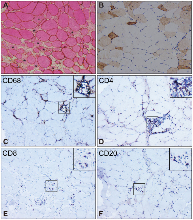
Figure 2.
Pathological characteristics of muscle biopsy in SRP-IMNM. (A), dystrophin combined with eosin staining reveals necrosis myofibers (asterisk). (B), immunohistochemical staining of Neural Cell Adhesion Molecule1 (NCAM1)/CD56 shows scattered myofiber regeneration. Inflammatory cell analysis demonstrates scattered or focal CD68+ macrophage infiltration and myophagocytosis (C), scattered CD4+ (D) and CD8+ T lymphocyte (E), and a few CD20+B lymphocytes (F). Magnification (A-F): 200x. These pictures are cited from our previous study (7, 63).

