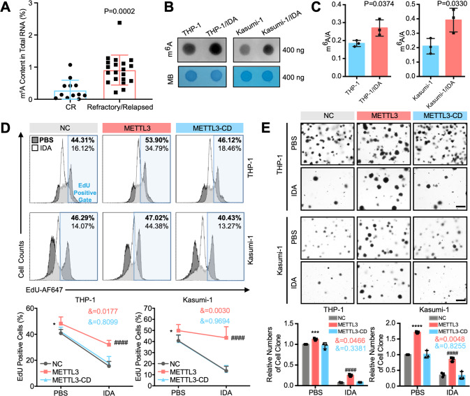
Fig. 2. METTL3-meditated m6A enhances the chemoresistance of AML cells.
A AML CD34+ cells from the patients shown in Fig. 1B were subjected to an m6A methylation quantification kit to assess global m6A changes (n = 13 for CR group and n = 20 for Refractory/Relapsed group). P < 0.05 was considered significant, t test. B, C m6A dot blot assays (B) and LC-MS/MS (C) for the detection of global m6A changes. MB, methylene blue staining (as a loading control). P < 0.05 was considered significant, t test. D EdU incorporation assay (upper) showing the percentage of AML cells that entered the proliferation cycle (EdU positive cells) with or without IDA pressure. Percentages after PBS treatment are shown in bold, and percentages after IDA treatment are shown in regular. Statistical analysis is shown in (lower). *P < 0.05, vs. the NC group with PBS treatment; ####P < 0.0001, vs. the NC group with IDA treatment; &, significant interaction effect; two-way ANOVA. E Colony-forming assays (upper). Bar, 500 μm. Statistical analysis is shown in (lower). ***P < 0.001, ****P < 0.0001, vs. the NC group with PBS treatment; ####P < 0.0001, vs. the NC group with IDA treatment; &, significant interaction effect; two-way ANOVA. n ≥ 3, mean ± SD values are shown for (A) and (C–E).

