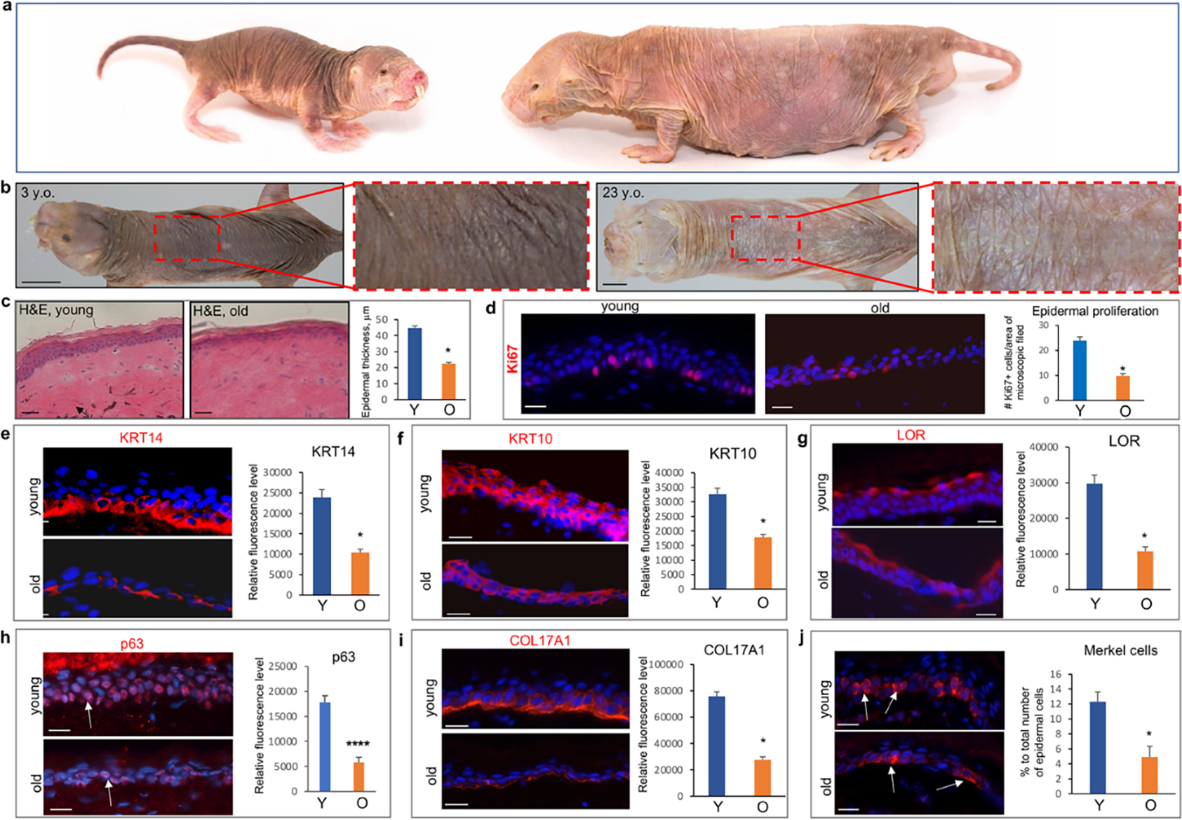
Figure 1. Visual appearance of the skin and age-related changes in the epidermis of NMRs.

a – Images of the skin in 3- and 23-year-old animals, note the more translucent and less pigmented dorsal skin in aged NMR. b – Hematoxylin/eosin staining of the young and old skin: presence of the dermal pigment (arrow) in young skin and significantly reduced epidermal thickness in old animals. c – Significant decrease in Ki67+ cells in aged versus young NMRs. d-f - Significantly decreased immunofluorescence intensity of K14 in the basal layer (d), K10 in the spinous layer (e), and Loricrin in the granular layer (f) in the epidermis of old NMRs. g - Significant decrease in the immunofluorescence intensity of p63 in epidermal keratinocytes of old versus young NMRs. h – Significant decline in the expression of COL17A1 in basal epidermal keratinocytes and dermal-epidermal basement membrane of old animals. i – Significant decrease in the number of KRT20+ Merkel cells in the epidermis of old NMRs (arrows). (mean ± SD, *p<0.05, Student’s t-test). Scale bars: 1b – 1 cm; 1c – 50 um; 1d-j – 25 um. Y – young animal, O – old animal.
