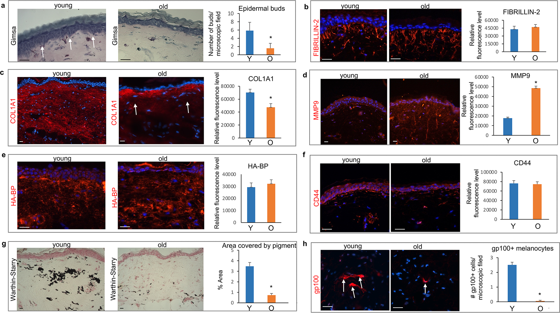
Figure 2. Age-associated changes in the NMR dermis.

(a) Significantly reduced number of epidermal buds elongating into the dermis (arrows) in the aged NMR skin. (b) Similar distribution of Fibrillin-2+ fibers in young and old NMRs. (c) COL1A1 is broadly expressed in the papillary and reticular dermis of young NMRs, while COL1A1 expression is significantly decreased in the reticular dermis in aged skin. (d) Significant increase in the MMP9 immunofluorescence intensity in the dermis of old NMRs compared to young animals. (e) No differences in the hyaluronan-binding protein (HA-BP) binding pattern or degree of binding between the dermis of young and old NMRs. (f) Similar expression of the HA receptor CD44 in the skin of young and old NMRs. (g) Warthin-Starry stain shows a dramatic decrease in the melanin containing areas in the dermis of old compared to young NMRs, which is accompanied by a significant decrease in the number of gp100+ pigment-producing dermal melanocytes in the dermis of aged animals (h, arrows). Mean ± SD, *p<0.05, Student’s t-test. Scale bars: 25 μm. Y – young animal, O – old animal.
