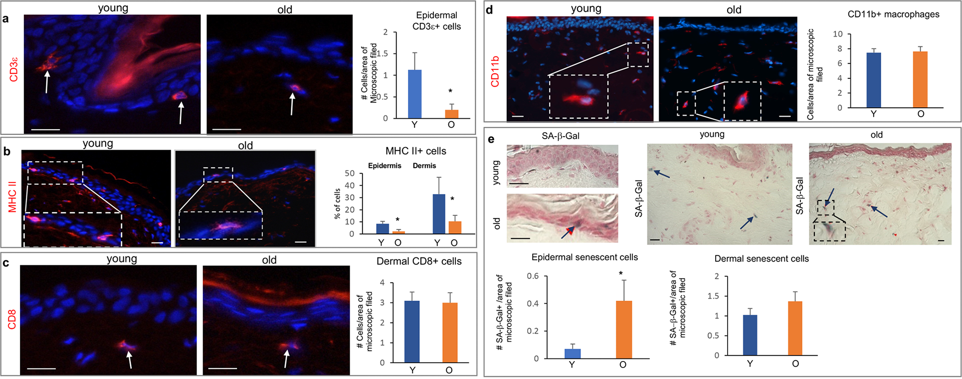
Figure 3. Aging-associated changes in the number of immune and senescent cells in the NMR skin.

(a) Significant decrease in the number of CD3ε+ T-cells in the epidermis of old NMRs. (b) Significant decrease in the number of MHC II+ cells detected by an anti-27E7 antibody in the epidermis and dermis of old NMRs. (c) No changes in the number of CD8+ cells in the dermis of aged versus young NMRs (arrows). (d) The number of dermal CD11b+ macrophages is similar in the young and old NMRs. (e) Significant increase in the number of senescent SA-β-gal+ cells (arrow) in the epidermis of old NMRs. A tendentious, but not significant, increase in the number of SA-β-gal+ cells (arrows) in the aged dermis (p=0.082). Mean ± SD, *p<0.05, Student’s t-test. Scale bars: 25 μm. Y – young animal, O – old animal.
