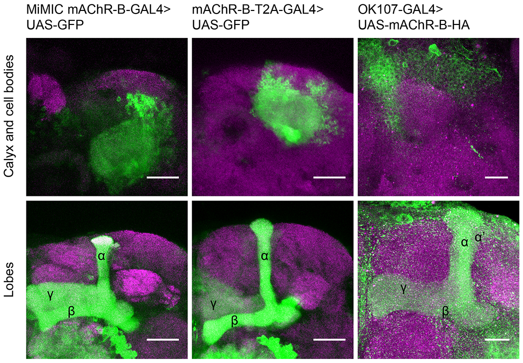
Figure 1: mAChR-B expression pattern.

Maximum intensity projection of confocal sections through the central brain of a fly carrying MiMIC-mAChR-B-GAL4 (left) or mAChR-B-T2A-GAL4 (middle), and UAS-GFP transgenes. MB αβ and γ lobes are clearly observed (bottom, 80 and 71 confocal sections respectively, 1 μm). Very weak GFP expression is observed in α’β’ lobes. As expected from soluble GFP labeling, the calyx is clearly observed (top, 44 and 24 confocal sections respectively, 1 μm). Right, the pan KC driver, OK107-GAL4 was used to overexpress UAS-mAChR-B-HA (with an HA tag). While KC axons at the MB lobes are clearly visible (bottom, 43 confocal sections, 0.5 μm), there is no expression at the calyx (compare to left and middle panels, 44 confocal sections, 0.5 μm) indicating mAChR-B is normally expressed in the axonal compartment.
